The Colossal Squid Eyeball — Inside the Largest Eye in History
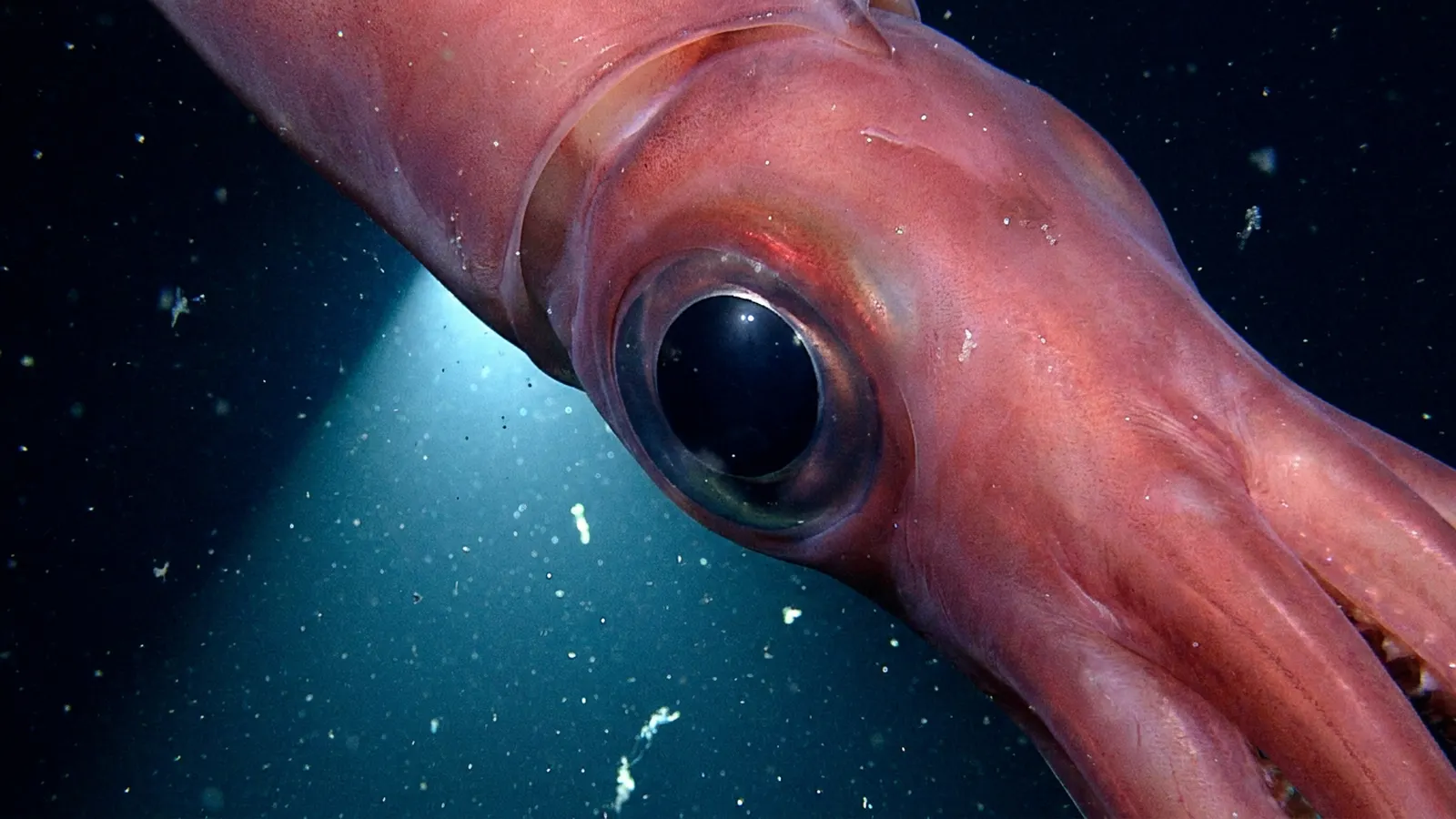
A colossal squid eyeball — 27 centimetres across — is a near-spherical structure containing several distinct layers. Outside to in: a tough sclera, a transparent cornea at the front, a muscular iris controlling a W-shaped pupil, a spheroidal crystalline lens roughly 10 cm across, a clear vitreous humour filling the interior, and a huge retina lining the back. Each layer is scaled up from a standard cephalopod eye, but the structural challenge of holding that much tissue together at depth is significant — especially given the pressure, cold, and density of Antarctic deep water.
An eyeball of this size is not just a scaled-up version of a smaller eye. It faces engineering problems that smaller eyes do not: structural support, optical correction across a huge lens, retinal wiring across a massive surface, and nourishment of tissue at distances where normal diffusion can’t keep up. The colossal squid’s eyeball solves all of these — it functions well enough to detect approaching sperm whales from 120 metres in near-darkness — but doing so requires a set of structural adaptations that distinguish it from the eye of any smaller cephalopod. Looking inside is a lesson in how biology builds a working camera-type eye at extreme scale.
The external structure — sclera, cornea, and mounting
A colossal squid eyeball sits in a socket of cartilage on the side of the head, held in place by the only significant cartilage structures in the animal’s body. The sclera — the tough outer shell of the eye — is thicker and more reinforced than in any other cephalopod.
On the outside, the eyeball has a white fibrous sclera, equivalent to the white of a human eye, that forms the outer wall of the sphere. In a colossal squid, the sclera is thick and reinforced with cartilage elements to maintain structural integrity against the animal’s own mantle contractions, against the pressure at depth, and against the forces of a tentacle struggle with prey. The front of the eye is transparent — the cornea — allowing light to enter while the rest of the sphere is opaque.
Mounted laterally on the squid’s head, the two eyeballs are oriented somewhat forward-facing, giving some binocular overlap in front while preserving wide peripheral vision. The muscular attachment allows each eye to move independently of the other, and a set of extraocular muscles can pivot the eyeball through a considerable range of angles. At the back of each eyeball, the optic nerve bundle emerges and connects to the squid’s optic lobes — the large, specialised processing centres that handle visual information in the central brain.
The lens — 10 cm of optical protein
At the optical centre of the eyeball sits a roughly spherical crystalline lens about 10 centimetres across. In a human eye, the lens is roughly 1 cm; in the colossal squid, the lens is ten times larger in each dimension.
Cephalopod lenses are built from densely-packed crystallin proteins arranged to create a refractive gradient — the centre of the lens has a slightly higher refractive index than the outer layers, helping correct the spherical aberration that would otherwise blur images produced by a large simple lens. Without this gradient, a 10-cm lens would produce unusable images; with it, the colossal squid’s eye achieves acceptable (though not sharp) focus across its entire visual field. The lens is supported by muscular elements that can move it slightly forward or back to focus on nearer or further objects, similar to the focusing mechanism in fish eyes. Unlike the human eye, which focuses by changing the shape of the lens, cephalopod eyes focus by moving the lens as a whole.
The scale of the lens is itself remarkable. A 10-cm spheroidal protein structure with precisely-controlled optical properties is a substantial engineering feat. It is larger than any known lens in any other animal. And it represents a significant fraction of the eyeball’s total volume — roughly 500 cubic centimetres, or half a litre of pure optical tissue.
The retina — millions of photoreceptors across a huge surface
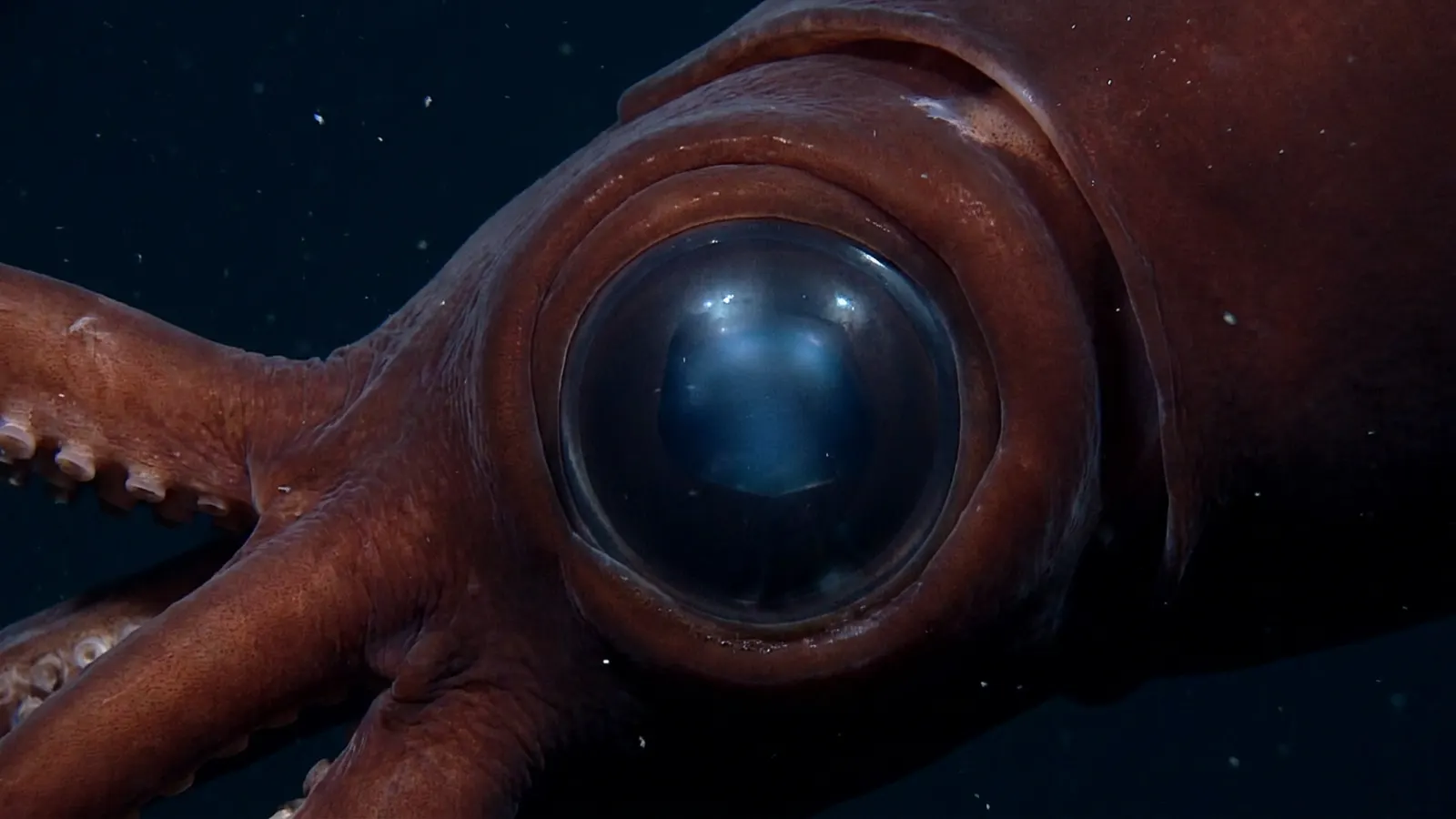
Lining the interior back wall of the eyeball is the retina, a photosensitive tissue with an enormous surface area. A single colossal squid retina covers roughly 2,000 square centimetres — the area of a small tablet screen — packed with tens of millions of photoreceptors.
Cephalopod retinas use a different photoreceptor cell type than vertebrate retinas, and the cells face the incoming light directly (rather than facing away, as in vertebrates). This means the colossal squid’s retina has no blind spot — nerve fibres run behind the photoreceptors and exit through the back of the eye without passing through the light-sensitive surface. It is an optically cleaner arrangement than a vertebrate retina, and across the massive area of a colossal squid eye, that cleanness matters.
The photoreceptor cells are tuned to blue wavelengths (around 470 nm), matching the dominant light in the deep sea. Each cell is sensitive enough to respond to small numbers of photons, and with tens of millions of such cells spread across the retina, the eye can integrate enough photons to detect extremely faint light sources. Resolution is poor compared to a human eye at the same distance — the cells are not as densely packed per unit area as in a sharp-focus eye — but sensitivity is extraordinary.
Comparison with the giant squid eyeball and others
The colossal squid’s eyeball is larger, more structurally reinforced, and positioned slightly more forward-facing than the giant squid’s. Structurally the two are similar, but every dimension is slightly larger in the colossal.
A giant squid eyeball is about 25 cm; a colossal squid’s is about 27 cm. The giant squid’s lens is about 9 cm; the colossal squid’s is about 10 cm. The retinal surface area is roughly proportional, meaning the colossal squid has somewhat more photoreceptors in total. Both eyeballs use the same camera-type architecture, the same W-shaped pupil, the same blue-tuned photoreceptors. The functional difference comes from the extra size — and the colossal squid’s slightly more forward-facing eye orientation, which gives some binocular depth perception the giant squid lacks.
Comparing to other large eyes: a blue whale eyeball is about 15 cm — smaller in every dimension. An ichthyosaur (Ophthalmosaurus) eyeball from the Jurassic period was estimated at 22 cm. An ostrich eyeball is about 5 cm. The colossal squid’s 27 cm is the winner against any known animal, alive or extinct. Within the animal kingdom, this eyeball is a record holder that has yet to be surpassed.
How big is a colossal squid eyeball?
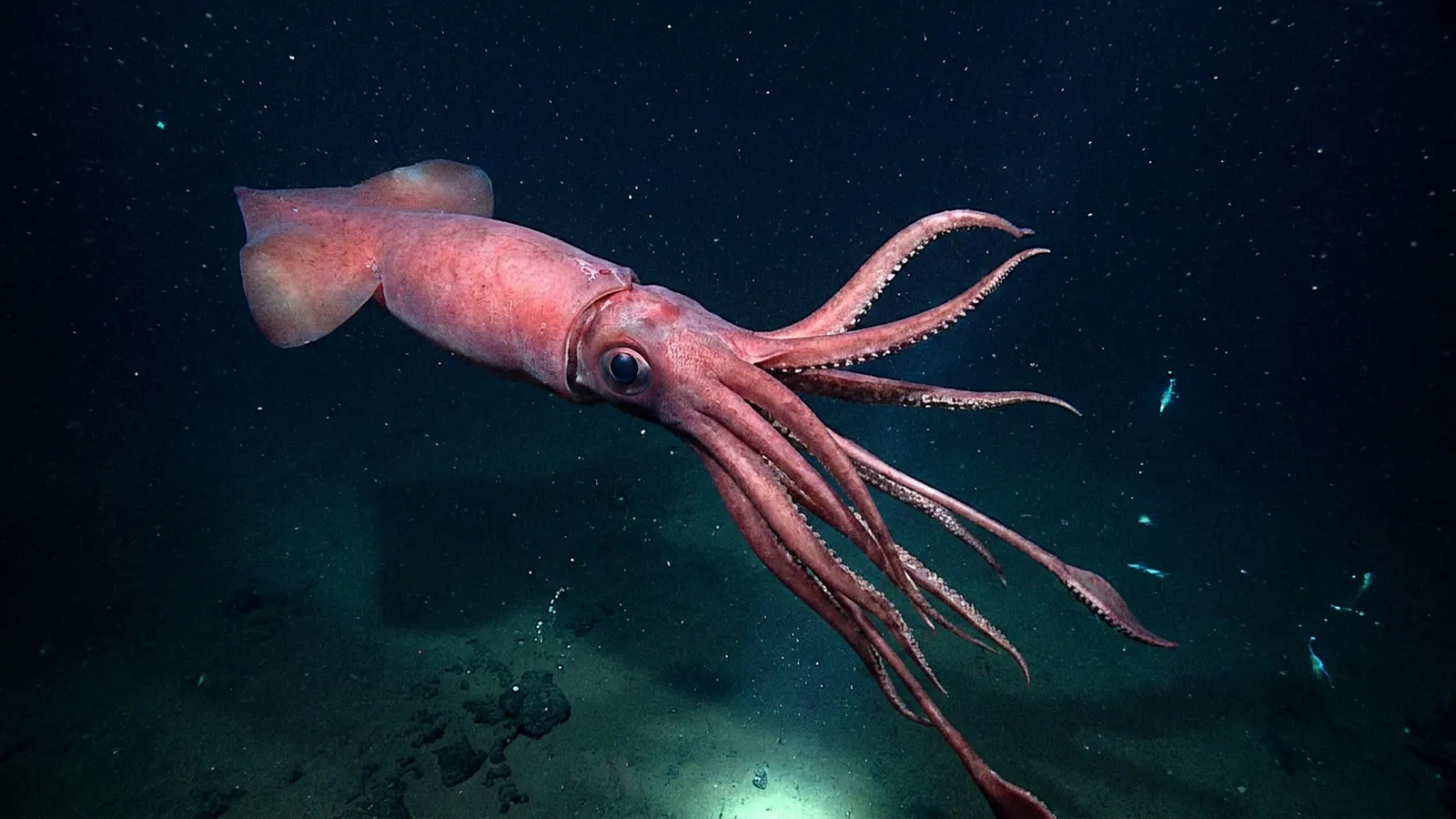
About 27 centimetres in diameter — the largest eyeball of any animal known to have lived. It contains a 10-cm lens, a retina covering about 2,000 square centimetres, and tens of millions of photoreceptor cells. The total eyeball volume is around 10 litres, weighing an estimated 3–5 kilograms.
What’s inside a colossal squid eyeball?
From outside in: a thick cartilage-reinforced sclera, a transparent cornea at the front, a muscular iris with a W-shaped pupil, a 10-cm spheroidal crystalline lens, a clear vitreous humour filling the interior, and a large retina lining the back wall. All components scaled up from the standard cephalopod eye.
How is a colossal squid eyeball different from a giant squid’s?
The colossal squid’s eyeball is about 2 cm larger in diameter (27 cm vs 25 cm). The internal components are proportionally larger, and the eye is positioned slightly more forward-facing, giving some binocular depth perception. Structurally, both eyes are camera-type with the same photoreceptor tuning and pupil shape, just at different scales.
How much does a colossal squid eyeball weigh?
No direct measurement has been published, but estimates based on volume and tissue density put each eyeball at roughly 3 to 5 kilograms. For a 500 kg animal with two eyeballs, that’s up to 10 kg of eye tissue — roughly 2% of total body mass concentrated in the visual system alone.
What does a colossal squid eyeball look like when preserved?
Intact preserved colossal squid eyeballs are extremely rare. The 2007 Ross Sea specimen’s eye was partially damaged during capture, but photographs show a pale, milky-white sphere about the size of a human head, with the lens visible as a darker rounded centre. Preserved specimens at Te Papa museum in New Zealand are among the few public displays of a colossal squid eyeball anywhere in the world.
Blane Perun has been exploring Earth’s oceans and marine life for over 25 years. Founder of TheSea.Org in 1999, underwater photographer, coral aquaculture pioneer, and explorer of 80+ countries.
About Blane Perun → More Squid Q&A →