Sponge Body Types — Asconoid, Syconoid and Leuconoid Sponges Explained
They are the three body plans of sponges, defined by where the pumping collar cells (choanocytes) sit. Asconoid sponges are simple tubes whose central cavity is lined directly with choanocytes; they stay small. Syconoid sponges fold the wall into radial canals lined with choanocytes. Leuconoid sponges have choanocytes in thousands of tiny chambers inside a thick body, with branching canals and many oscula. The leuconoid plan is by far the most common and is the only one that allows sponges to grow large.
Every sponge solves the same problem: how to move as much water as possible past as many collar cells as possible, so it can catch enough food and oxygen to live. The three classic body types, asconoid, syconoid and leuconoid, are three solutions to that problem, each more elaborate than the last. They are a staple of biology classes because they show neatly how a simple design can be scaled up by folding, a trick that turns up again and again in biology, from the lining of our intestines to the gills of fish.
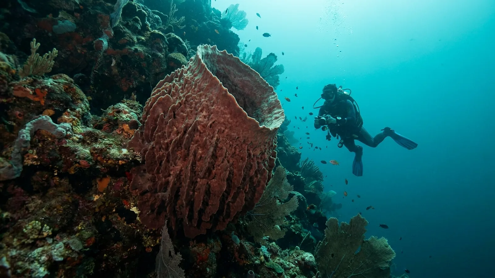
On the Caribbean walls I’ve dived, the biggest sponges are hard to miss, and they are the end point of this story. The giant barrel sponges on Cozumel’s walls, which grow large in the island’s plankton-rich current, are leuconoid sponges, and they could not exist with either of the simpler designs. Understanding why is a good way to understand how sponges work.
The problem every sponge has to solve
A sponge feeds by pumping water. The pump is the choanocyte, or collar cell, and the more choanocyte surface a sponge has relative to its volume, the more food it can gather.
Each choanocyte has a whip-like flagellum that drives water and a collar of microvilli that catches bacteria and tiny particles. If a sponge simply grows bigger without changing shape, its volume increases much faster than the surface area of the cavity lined with choanocytes. A large hollow tube would end up with a huge volume of water in the middle that its few collar cells could never move or filter efficiently. The solution is to increase the choanocyte surface by folding it. That folding is what distinguishes the three body types. See our labeled sponge diagram if you need a refresher on the basic parts.
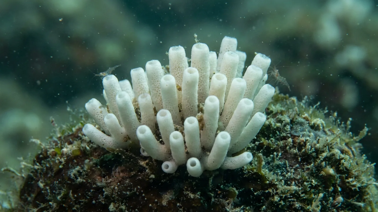
Asconoid sponges: the simple tube
Asconoid sponges are the simplest. Each is a thin-walled tube or vase whose central cavity, the spongocoel, is lined directly with choanocytes.
Water enters through pores (ostia) that pass straight through the thin wall, often each through a single tube-shaped porocyte cell. It is driven by the choanocytes lining the cavity and leaves through a single osculum at the top. Because the choanocyte layer is just a single lining around a large hollow space, asconoid sponges are inefficient pumps, and they never grow large; most are only a few millimetres to a few centimetres across. Many grow as clusters of small, branching tubes. The asconoid form is found only in some calcareous sponges, such as Leucosolenia and Clathrina, which often look like delicate white or pale yellow lacework on rocks and in caves. Our page on calcareous sponges covers this group.
Syconoid sponges: folding the wall
Syconoid sponges increase their pumping surface by folding the body wall outward into many finger-like radial canals.
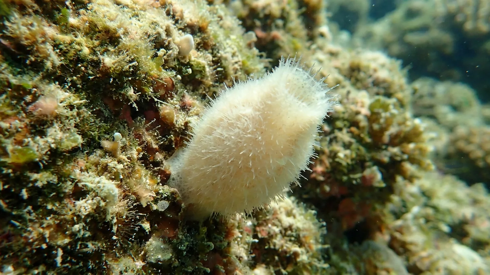
In a syconoid sponge, the choanocytes no longer line the central cavity. Instead they line the radial canals, which radiate out from the spongocoel like the spokes of a wheel. Between the radial canals run incurrent canals, open to the outside. Water enters through surface pores into the incurrent canals, passes through tiny openings called prosopyles into the radial canals, where the choanocytes pump and filter it, then flows through openings called apopyles into the spongocoel and out of the osculum. The result is a thicker wall with much more choanocyte surface. The classic example is Sycon, sometimes called Scypha, a small, bristly, vase-shaped calcareous sponge often used in biology labs. Syconoid sponges can grow larger than asconoid ones but are still usually small.
Leuconoid sponges: chambers and canals
Leuconoid sponges take the folding much further. The choanocytes are packed into thousands or millions of tiny, rounded flagellated chambers scattered through a thick body.
Water enters through surface pores into a branching network of incurrent canals, which lead to the choanocyte chambers. From there it flows into excurrent canals that merge into larger channels and finally exit through one or many oscula. The central spongocoel is reduced to a set of excurrent canals or disappears altogether. Because the chambers are tiny, water slows down as it passes the collars, giving the choanocytes more time to catch food, and then speeds up again in the narrowing excurrent canals, shooting out of the oscula with enough force to carry waste away. This design can be scaled up almost indefinitely. Nearly all demosponges, which make up most of the world’s sponges, are leuconoid, including bath sponges, barrel sponges, tube sponges and most other reef sponges, as are many calcareous and glass sponges.
| Feature | Asconoid | Syconoid | Leuconoid |
|---|---|---|---|
| Where choanocytes are | Lining the spongocoel | Lining radial canals | In many small chambers |
| Spongocoel | Large, central | Present | Reduced or absent |
| Body wall | Thin | Folded, thicker | Thick, with canal network |
| Oscula | One per tube | Usually one | Often many |
| Typical size | Millimetres to a few cm | A few cm | Up to metres |
| Pumping efficiency | Low | Medium | High |
| Found in | Some Calcarea | Some Calcarea | Most demosponges; many Calcarea and glass sponges |
| Example | Leucosolenia | Sycon | Giant barrel sponge, bath sponge |
Why big sponges are always leuconoid
Every large sponge you will see on a reef is leuconoid, and the reason is simple geometry.
By packing choanocytes into countless small chambers, a leuconoid sponge keeps the ratio of pumping surface to body volume high no matter how big it grows. A giant barrel sponge can grow taller than a diver and is estimated to live for centuries; see our page on the giant barrel sponge. It pumps enormous quantities of water every day, filtering bacteria and particles out of the reef water and playing a large role in reef nutrient cycling. A simple asconoid tube scaled up to that size would be a hollow cylinder full of stagnant water. The leuconoid design is also the most flexible: it can take the form of crusts, tubes, vases, branching trees or massive mounds, which is why sponges come in such a variety of shapes. On the walls of Cozumel, Cayman and Roatan, the sponges you see in every size and colour are all variations on the leuconoid theme.
Seeing the body types for yourself
You do not need a microscope to see the difference between a simple sponge and a leuconoid one, though it helps.
On a reef, look for the oscula. A small, pale, tube-shaped calcareous sponge growing in a shaded crevice will often have a single opening at the end of each little tube, the hallmark of the simpler body plans. A large leuconoid sponge, by contrast, may be covered in dozens or hundreds of oscula of different sizes, each the outlet for its own network of canals. Tube sponges show this well: each tube has a large osculum at the top, but the tube wall is thick and riddled with canals, not a thin asconoid sheet. If you hover quietly near a big sponge and watch for fine particles in the water, you can sometimes see the outflow from an osculum as a faint shimmer or a stream of drifting particles.
In a reef aquarium, small calcareous sponges often appear on their own in dark places such as overflow boxes, sumps and the undersides of rock. Many of them are tiny white tubes or bristly vases that look very like the Sycon and Leucosolenia pictured in biology textbooks. They are usually harmless filter feeders and a sign of a healthy system with plenty of fine food in the water, as covered in our page on sponges in reef tanks. For students, a sample of one of these under a simple microscope, or a cross-section of a Sycon from a biological supplier, makes the canal system easy to see.
Are body types a sign of evolution?
It is tempting to see the three types as steps in an evolutionary ladder, but biologists are cautious about that.
The three body plans are best treated as grades of organisation rather than family groups. They do not map neatly onto sponge classes: asconoid and syconoid forms are found only in some calcareous sponges, while leuconoid forms occur in all four classes. Molecular studies suggest that the leuconoid plan may have arisen more than once, and that some calcareous sponges have changed body type during their evolution. Interestingly, many leuconoid sponges pass through a simpler stage as they develop from a larva into an adult, and some textbooks describe an intermediate, sylleibid, arrangement as well. The neat three-step sequence is a useful teaching tool, but real sponge evolution is messier, as it usually is. For the four sponge classes, see sea sponge species.
What is an asconoid sponge?
The simplest sponge body plan: a thin-walled tube whose central cavity is lined directly with choanocytes, with water exiting through one osculum.
What is the difference between syconoid and leuconoid sponges?
Syconoid sponges have choanocytes lining radial canals around a central cavity; leuconoid sponges have choanocytes in many small chambers within a thick body, with branching canals and often many oscula.
Which sponge body type is most common?
Leuconoid. Nearly all demosponges, which make up most sponge species, are leuconoid.
Why are asconoid sponges small?
Their single layer of choanocytes cannot pump and filter enough water to support a large body volume.
What is an example of a syconoid sponge?
Sycon (also called Scypha), a small vase-shaped calcareous sponge.
Is a bath sponge leuconoid?
Yes. Commercial bath sponges are demosponges with a leuconoid body plan and a skeleton of spongin fibres.
- Brusca, R. C., Moore, W. & Shuster, S. M. (2016). Invertebrates, 3rd ed. Sinauer Associates.
- Leys, S. P. & Hill, A. (2012). The physiology and molecular biology of sponge tissues. Advances in Marine Biology 62: 1–56.
- Manuel, M. et al. (2003). Phylogeny and evolution of calcareous sponges. Systematic Biology 52: 311–333.
- World Porifera Database (WoRMS).
Blane Perun has been exploring Earth’s oceans and marine life for over 25 years. Founder of TheSea.Org in 1999, underwater photographer, coral aquaculture pioneer, and explorer of 80+ countries.
About Blane Perun → More on Sponges →