Yellow Goniopora — A Sunny Color That Depends on Your Spectrum
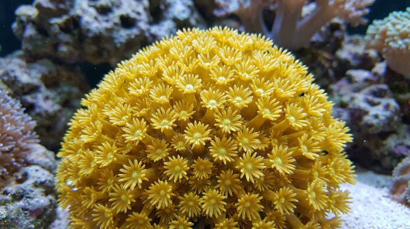
Yellow Goniopora is a trade-name color form of flowerpot coral, less common than green. Yellow tones usually come from green-yellow fluorescent proteins in the coral tissue, or a blend of green and red ones, sometimes mixed with tan zooxanthellae, and colonies often show yellow tentacles around green or white mouths. Because fluorescence depends on the light hitting it, yellow can look lemon under blue-heavy lighting and tan under white light. Care is standard: moderate, stable light; moderate random flow; stable parameters; and fine-particle feeding.
Yellow is one of the trickier Goniopora colors to photograph honestly, because it shifts so much with lighting. Judge it in person under the light you’ll use at home.
What makes Goniopora yellow?
Reef animals make fluorescent proteins in a range of emission colors, from cyan through green to yellow and red, although true yellow emitters are documented mainly from zoanthids rather than stony corals. A yellow Goniopora is usually showing a long-wavelength green fluorescent protein, or a mix of green and red, with the coral’s brown-gold zooxanthellae adding warmth. The balance between pigment and algae depends on light and nutrients, so yellow is often a moving target.
| Condition | Effect on yellow |
|---|---|
| Blue-rich lighting | Brighter lemon-yellow fluorescence |
| White-heavy lighting | More tan or gold |
| Low light / high nutrients | Browner |
| Sudden light increase | Pale or bleached |
Is yellow Goniopora hard to keep?
It needs standard Goniopora care, and color doesn’t change the coral’s basic needs. Choose aquacultured colonies when you can, and inspect wild colonies carefully for pests, recent frag wounds and exposed white skeleton.
Why is my yellow Goniopora turning brown or pale?
Brown usually means less light or more nutrients. Pale or white suggests too much light or other stress. Check recent changes: new lights, a moved colony, a temperature swing or a nutrient spike. Correct slowly and give it weeks.
Is it Goniopora or Alveopora?
Yellow is common in both. Count the tentacles on an open polyp: 24 for Goniopora, 12 for Alveopora. More in Alveopora vs Goniopora.
Where should yellow Goniopora be placed?
In the middle of the tank or on a well-lit sand bed, in moderate random flow. See Goniopora placement, care and feeding. For the closely related green form, see green Goniopora.
What lighting shows yellow Goniopora best?
Balanced reef lighting with a moderate blue component usually shows yellow most naturally. Very blue-heavy lighting makes the fluorescence pop but can push yellow toward green, while daylight-heavy lighting reveals more of the tan algae beneath. If you photograph the colony, use a yellow or orange lens filter or a white-balanced camera setting under blue light; otherwise the picture will exaggerate the glow. When you change fixtures, run the new light at reduced intensity for a few weeks and raise it gradually, watching polyp extension as well as color.
- Balanced spectrum for natural yellow.
- Extra blue for fluorescence, at the cost of accuracy.
- White-heavy light reveals tan undertones.
- Ramp new lights over weeks.
Is the yellow made by the coral or by its algae?
Both partners can contribute, and the two kinds of colour behave differently. The coral animal makes GFP-like proteins in its own tissue, and some of those proteins fluoresce, turning absorbed blue light into longer wavelengths. The single-celled algae living inside the coral’s gut lining contribute ordinary pigments that absorb light without re-emitting it as visible colour. A colony that looks yellow in a shop is usually showing a blend of the two, and the proportions decide whether it reads as lemon, gold, mustard or khaki.
There is a simple way to see which partner is doing most of the work. Switch the tank to blue actinic light only. A surface that is yellow purely because it reflects yellow light needs yellow wavelengths to reflect, and a blue-only lamp supplies almost none, so algal or reflected yellow turns dull brown or nearly black. Yellow that survives under actinic light, or even brightens, is being generated inside the tissue by fluorescence. Most yellow Goniopora fall somewhere between the two extremes, glowing on the tentacle tips while the polyp bases darken.
What pigments give the algae their golden colour?
The algae belong to the dinoflagellate family Symbiodiniaceae, long lumped together as zooxanthellae. That old name is itself a description of their colour: Roth’s 2014 review of coral photobiology notes that it comes from zoo, animal, and xanth, yellow. In 2018 LaJeunesse and colleagues split the single genus Symbiodinium into several genera within the family and dated its earliest diversification to roughly 160 million years ago, in the Mesozoic.
Their light-harvesting pigments are chlorophyll a, chlorophyll c2 and a carotenoid called peridinin. According to the absorption values compiled by Roth, chlorophyll a absorbs most strongly around 435 to 440 nanometres and again in the red near 670 to 680 nanometres, chlorophyll c2 absorbs around 450 to 460 nanometres, and peridinin peaks around 478 to 500 nanometres. Peridinin widens the band of usable light because it absorbs broadly from roughly 450 to 550 nanometres, deep into the blue-green.
The same algae also make beta-carotene and two further xanthophylls, diadinoxanthin and diatoxanthin. Because these carotenoids soak up blue and blue-green light, what is left to reflect back to the eye is weighted toward yellow, orange and red. Packed densely enough, the result is the familiar brown of an ordinary coral. Spread more thinly in tissue that also carries fluorescent protein, the same pigments read as a warm gold under the bright tentacles.
How many algal cells sit in a square centimetre of coral?
More than most people imagine. Roth’s review gives typical densities of 1 to 4 million algal cells per square centimetre of coral surface, with values as high as 8 million recorded, and notes that reef-building corals usually hold one or two algal cells in each host gut cell. The coral is thought to keep that population in check largely by limiting the nitrogen available to its symbionts, although the details are still not well understood.
Those numbers explain why a yellow colony can drift toward brown when nitrate and phosphate climb. More available nitrogen lets the algae multiply and pack more pigment, and every extra cell adds another layer of golden-brown filter over the coral’s own fluorescent proteins. The reverse is also true. A colony that suddenly looks pale and lemon-bright may have lost part of its algal population, letting the host colour show through more strongly.
What is the xanthophyll cycle, and can it shift colour?
It is a fast protective switch inside the algae. Within minutes of exposure to strong light, the algae convert diadinoxanthin into diatoxanthin, and the change reverses again when light becomes limiting. A higher proportion of diatoxanthin is associated with protection of the photosynthetic machinery from excess light energy.
In practice the cycle is too quick and too subtle to explain the colour changes hobbyists notice over weeks. Its value is as a reminder that the algal half of a yellow Goniopora is not a fixed paint layer. It is a living system that adjusts within minutes to light and within days to light and nutrient conditions. Roth’s review reports that algal density in corals can acclimate to a new light intensity within 15 days, and that the amount of chlorophyll and peridinin per cell is adjusted on a similar timescale while the ratios between pigments stay much the same.
Do stony corals make true yellow fluorescent proteins?
Rarely, if at all, on current evidence. The first yellow-emitting member of the GFP family from a reef animal, zFP538, was cloned by Matz and colleagues in 1999 from a button polyp of the genus Zoanthus. That is a zoanthid, not a stony coral. Its name records its emission peak at 538 nanometres, which sits between typical green and orange-red coral proteins.
Crystal structures published by Remington and colleagues in 2005 showed that zFP538 carries an unusual three-ring chromophore. A lysine in the protein chain forms an extra ring and cuts the backbone, adding one more double bond to the light-absorbing system than GFP has. That longer system shifts both absorption and emission toward lower energy, which is why the protein glows yellow rather than green. A 2005 study by Zubova and colleagues found that zFP538 also forms large aggregates in solution, and that its brightness depends on that aggregation.
For the stony corals themselves, the 2008 survey by Alieva and colleagues added 40 new GFP-like proteins and described the typical coral palette as cyan, green and red fluorescent colours plus non-fluorescent purple-blue chromoproteins. The emission ranges collected in Roth’s 2014 table run from about 497 to 525 nanometres for coral green proteins and from about 572 to 609 nanometres for red ones, leaving a gap where a pure yellow emitter would sit.
So where does a fluorescent yellow come from?
Two explanations fit the evidence. The first is a green protein at the long-wavelength end of its range, emitting near 520 to 525 nanometres, which the eye reads as yellow-green and which looks yellower still beside a darker, browner background. The second is a mixture of colours. Human vision combines green and red light into a single sensation of yellow, so tissue carrying both green and red fluorescent proteins in the right balance can appear yellow or gold even though neither protein emits yellow.
Those explanations are hypotheses for any given colony rather than measurements. Nobody has published the fluorescent protein complement of a yellow Goniopora sold in the aquarium trade, and spectral measurement of the tentacles would be the only way to tell a long-wavelength green from a green-plus-red mix. What can be said with confidence is that the yellow is not a single fixed pigment, which is why it moves so readily with light and nutrients.
Does the skeleton change how the colour looks?
Yes. Light that is not absorbed by the tissue or its algae is mostly reflected by the white aragonite skeleton as diffuse reflectance, and some of it enters the porous skeleton and re-emerges elsewhere. Roth’s review explains that this multiple scattering sends light back through the tissue, amplifying the light the algae receive.
The same effect brightens a coral’s colours. Pigments and fluorescent proteins get a second pass at the light, so a thin tissue layer over a bright skeleton can look more saturated than the pigment concentration alone would suggest. Bollati and colleagues showed in 2020 that this reflected light is also part of the feedback that drives colourful bleaching: once algae are lost, more light bounces around in the tissue and the host responds by making more protective pigment. A yellow Goniopora whose polyps retract for long periods exposes more skeleton to the light, which is one reason a colony’s colour can look different from week to week.
How do scientists record coral colour?
Field programmes avoid words like yellow because names mean different things to different observers. The CoralWatch Coral Health Chart from the University of Queensland uses printed colour squares representing the most common reef colours, each on a 6-point brightness scale. Observers compare the lightest and darkest parts of a colony with the chart and record codes such as C2 or D4. CoralWatch explains that the brightness levels correspond to differing concentrations of symbiotic algae, because coral colour correlates with chlorophyll content and algal density.
Laboratory work goes further and measures reflectance spectra. A 2026 study by Watty and colleagues in Coral Reefs calibrated a reflectance index called NDVI against measured chlorophyll a in three coral species and found a strong but non-linear relationship. They attributed the curve at the healthy end to pigment self-shading, and the curve at the bleached end to light backscattered from the increasingly exposed skeleton, and recommended species-specific calibrations. The lesson for a hobbyist is the same at smaller scale: colour is a useful health indicator, but only when it is compared like with like.
How can you track a yellow colony’s colour at home?
Borrow the field methods. Photograph the colony once a week from the same position, at the same time in the light cycle, with the same camera settings and white balance locked. Include a fixed reference in the frame, such as a small white or grey card mounted outside the glass, so that changes in the image can be separated from changes in the camera. A printed colour chart works for this purpose too.
Take two pictures each time: one under the normal display spectrum and one under actinic light alone. The first shows the combined appearance. The second isolates the fluorescent part, because reflected algal colour drops away under blue light. If the actinic image stays steady while the white-light image browns, the algae are increasing. If the actinic image brightens sharply while the white-light image pales, check your temperature log, because that pattern can mark the start of colourful bleaching rather than improving health.
Does the yellow tell you anything about health?
On its own, not much. A deep, even yellow on fully extended polyps is consistent with a healthy colony, but the same shade appears on colonies with very different algal loads and lighting histories. What carries information is change. Sudden paling, a shift toward intense neon tones a few weeks after a temperature spike, or tissue drawing back from the skeleton all deserve attention, whatever the starting colour. A slow drift toward gold or tan after a lighting change is far more often acclimation than illness.
Is yellow Goniopora rare?
It is less common than green but regularly available.
Why does yellow Goniopora look different at home?
Fluorescent colors change with lighting spectrum.
Why is my yellow Goniopora turning brown?
Lower light or higher nutrients.
Is yellow Goniopora a species?
No, it is a color form.
How do I care for yellow Goniopora?
Moderate light, random flow, stable water and fine-food feeding.
Does yellow Goniopora glow?
Its fluorescent pigments can glow under actinic light.
Can yellow Goniopora turn green?
Color can shift with light and nutrients over weeks.
- Roth, M.S. (2014). The engine of the reef: photobiology of the coral–algal symbiosis. Frontiers in Microbiology 5: 422. doi:10.3389/fmicb.2014.00422
- LaJeunesse, T.C. et al. (2018). Systematic revision of Symbiodiniaceae highlights the antiquity and diversity of coral endosymbionts. Current Biology 28: 2570–2580. doi:10.1016/j.cub.2018.07.008
- Matz, M.V. et al. (1999). Fluorescent proteins from nonbioluminescent Anthozoa species. Nature Biotechnology 17: 969–973. doi:10.1038/13657
- Remington, S.J. et al. (2005). zFP538, a yellow-fluorescent protein from Zoanthus, contains a novel three-ring chromophore. Biochemistry 44. doi:10.1021/bi048383r
- Zubova, N.N. et al. (2005). Brightness of yellow fluorescent protein from coral (zFP538) depends on aggregation. Biochemistry. doi:10.1021/bi048274c
- Alieva, N.O. et al. (2008). Diversity and evolution of coral fluorescent proteins. PLoS ONE 3: e2680. doi:10.1371/journal.pone.0002680
- Bollati, E. et al. (2020). Optical feedback loop involving dinoflagellate symbiont and scleractinian host drives colorful coral bleaching. Current Biology 30: 2433–2445. doi:10.1016/j.cub.2020.04.055
- Watty, K., Schoepf, V., Johnson, K.W., Littke, S. & van der Zande, R.M. (2026). Using reflectance to measure chlorophyll a in corals: calibration and implications of skeletal optical properties. Coral Reefs. doi:10.1007/s00338-026-02831-0
- CoralWatch, University of Queensland: Coral Health Chart and FAQs (coralwatch.org, accessed November 2026).
- Reef Builders (2025): Introduction to Goniopora care.
Blane Perun has been exploring Earth’s oceans and marine life for over 25 years. Founder of TheSea.Org in 1999, underwater photographer, coral aquaculture pioneer, and explorer of 80+ countries.
About Blane Perun →