Does a Sea Sponge Have a Brain? — No Brain, No Nerves, Yet It Still Reacts
No. Sea sponges have no brain, no nerve cells and no nervous system of any kind. They also lack muscles and organs. Even so, sponges are not inert: they sense touch, sediment and chemicals, can slowly contract their bodies, and some perform a coordinated “sneeze” to flush out their canals. They do this with chemical signals passed between cells and, in glass sponges, fast electrical signals through their tissue.
A sponge is a loosely organised colony of specialised cells. Each cell responds to its own surroundings, and cells communicate by releasing and detecting chemicals, much as cells in our own bodies do. Because sponges don’t need to hunt, flee or coordinate complex movement, slow chemical signalling is enough. See sea sponge anatomy for the cell types involved.
No brain, no nerves: what a sponge has instead
A sponge is a body built around a water pump, run by a handful of specialised cell types and no central controller.
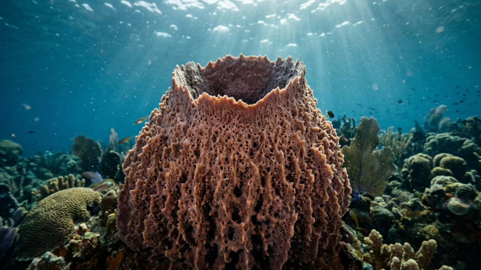
Sponges belong to the phylum Porifera, the “pore bearers”. A 2012 census of the World Porifera Database counted 8,553 valid living species, about 83% of them in the class Demospongiae, and new species are described every year. Whatever their shape, from encrusting crusts to vases taller than a diver, they share one plan. Water enters through thousands of small pores, flows through a branching system of canals and chambers, and leaves through one or more larger openings called oscula. Along the way, bacteria and fine organic particles are filtered out and eaten.
The work is divided among a few cell types rather than organs. Flattened pinacocytes form the outer skin and line the canals. Porocytes form the inlet pores in some groups. Choanocytes, or collar cells, line the feeding chambers; each beats a single flagellum to drive water and traps food on a collar of tiny projections. Between these layers lies the mesohyl, a jelly-like middle layer where amoeboid cells crawl about, digest food, carry nutrients, build the skeleton of spicules or spongin fibres and, when needed, turn into other cell types. A whole-body single-cell RNA study of the freshwater sponge Spongilla lacustris, published in Science in 2021, identified 18 distinct cell types. None of them is a neuron and none is a true muscle cell.
That is the core of the answer. A brain is a concentration of nerve cells that collects signals, processes them and sends out commands. Sponges have no nerve cells to concentrate, no nerve fibres to carry messages and no synapses where nerve cells pass signals to each other. There is no head end and, in most species, no fixed front or back. There is nowhere for a brain to be and nothing for it to do in the way it does in a fish or an octopus.
How do sponges sense anything without nerves?
Through cells that act as their own sensors, including antenna-like cilia that seem to measure water flow.
The clearest evidence comes from the osculum, the chimney through which water leaves the sponge. In 2014, Danielle Ludeman, Sally Leys and colleagues reported in BMC Evolutionary Biology that the cells lining the inner surface of the osculum in freshwater demosponges carry short cilia. Under the electron microscope these cilia lack the central pair of microtubules found in cilia that beat, and high-speed video confirmed that they do not move. In other animals, including humans, non-motile “primary” cilia work as cellular antennae that detect fluid flow and chemical signals.
The researchers tested whether the sponge cilia did the same. Drugs that block the ion channels used by sensory cilia in other animals reduced or abolished the sponge’s whole-body contractions. Stripping the cilia off chemically, or cutting off the entire osculum, also stopped the contractions, and in every case the effect was reversible once the treatment ended. Sponge transcriptomes turned out to contain several transient receptor potential (TRP) channel genes, a family that includes the PKD channels linked to flow sensing in other animals. The authors concluded that the osculum works as a simple sensory organ: the cilia appear to register how fast water is flowing and whether the canals are clogging, and the sponge responds by changing the shape of its canal system.
Sponges also respond to touch, to sediment in the water and to particular chemicals, as the experiments below show. What they lack is a place where all this information is gathered and weighed. Each region reacts locally, and its reaction spreads to neighbouring tissue.
The sponge “sneeze”: coordination without a coordinator
When their canals clog, many sponges carry out a slow, orderly sequence of contractions that flushes the waste out.
The best-studied example is Ephydatia muelleri, a freshwater sponge that is easy to grow in the laboratory from overwintering buds called gemmules. In a 2007 paper in the Journal of Experimental Biology, Glen Elliott and Sally Leys filmed the response that follows a mechanical stimulus such as a shake or a burst of sediment. First, the tip of the osculum constricts and the excurrent canals swell. Fields of inlet pores on the sponge’s upper surface close in unison. Then the inner tissue, the outer skin and the osculum contract, squeezing water and clumps of waste out of the canal system. Finally everything relaxes back to its normal state. A single stimulus typically set off three of these “inflation-contraction” cycles in a row.
The waves are slow and their speed depends on the tissue. Elliott and Leys measured 0.3–1 micrometre per second in peripheral canals, 1–4 micrometres per second in central canals and 6–122 micrometres per second in the osculum. Even the fastest rate is thousands of times slower than a typical nerve impulse. Cells crawling in the mesohyl paused as a wave of contraction passed them, a hint that a chemical signal was moving between cells. The contractile machinery is also unusual: bundles of actin filaments run through the pinacocytes of the outer skin and link up from cell to cell through dense plaques, forming continuous tracts across the whole animal. The sponge is, in effect, using its skin as both sensor and muscle.
How long does a sneeze take? It depends on size. Small laboratory sponges complete the sequence in under an hour. In the wild, larger sponges are far slower, as a four-year camera study in British Columbia showed. The fact that the sequence is the same every time, ordered and repeatable, is what convinced researchers that sponges show genuine coordinated behaviour, not just random twitches.
Which chemical messengers do sponges use?
Many of the same small molecules that serve as neurotransmitters in animals with nerves.
In a 2010 follow-up, Elliott and Leys showed that glutamate, an amino acid that is the main excitatory neurotransmitter in the human brain, triggers the full sneeze sequence in Ephydatia, and that the effect grows with the dose. Drugs that block metabotropic glutamate receptors stopped glutamate-triggered contractions. GABA, the main inhibitory neurotransmitter in vertebrates, did the opposite and suppressed contractions. The team found pools of glutamate, glutamine and GABA in the tissue, consistent with a working signalling system. They also located nitric oxide synthase, the enzyme that makes the gas nitric oxide, in cells of the osculum and outer skin.
A 2025 study from the same laboratory added ATP to the list. ATP is the cell’s energy currency, but in many animals it is also released as a signal between nerve cells and acts on P2X receptors. In Ephydatia, ATP made the excurrent canals expand, and its breakdown product ADP triggered complete contractions. Blocking P2X receptors prevented contractions triggered by both ATP and glutamate, which suggests ATP acts downstream of glutamate. The sponge genome holds two P2X receptor genes.
These molecules are not nerve signals in a sponge. They diffuse through the tissue and the water in the canals and act on nearby cells. That is far slower and less targeted than a nerve impulse jumping across a synapse, but for an animal that only needs to adjust a pump over minutes or hours it is enough. It also shows that much of the chemistry of the nervous system existed before nerves did.
Rhythms in a sponge that never stops moving
Some sponges contract on a regular beat, and that beat changes between day and night.
Tethya wilhelma, a small ball-shaped demosponge first discovered in the aquarium of the Wilhelma zoo in Stuttgart, has become a model for sponge behaviour because it contracts rhythmically. Time-lapse filming by Michael Nickel, published in 2004, showed that it can shrink its body volume by up to 73.3% in a regular contraction. Each cycle follows the same four phases. Over more than a week of recording, the cycles were significantly longer during the dark hours, showing a day-night periodicity. Contractions started locally and spread over the surface at about 12.5 micrometres per second. When two pieces of the same clone were fused, each kept its own rhythm for several days until the fused tissue became a single sponge with one synchronised rhythm.
In 2006, Kornelia Ellwanger and Nickel tested chemical messengers on the same species. Caffeine, glycine, serotonin, nitric oxide and cyclic AMP all induced contractions. Adrenaline and nitric oxide slowed the natural rhythm, while caffeine and glycine sped it up. All of the effects disappeared when the substances were washed out. The result again points to a chemical coordination system built from molecules that later animals recruited for their nervous systems.
Glass sponges: electrical signals without nerves
One group of sponges does send electrical impulses, though not through nerve cells.
Glass sponges, class Hexactinellida, are mostly deep-water animals with skeletons of silica. Unlike other sponges, much of their tissue is a syncytium: a continuous web of cytoplasm with many nuclei and few cell boundaries. In 1999, Sally Leys, George Mackie and Robert Meech recorded electrical impulses in the glass sponge Rhabdocalyptus dawsoni from British Columbia. Small electric shocks, a touch, or particles added to the incoming water each set off an all-or-none impulse that spread through the tissue at about 0.27 centimetres per second.
A thermistor flow meter showed that water flow through the sponge stopped after the impulse passed, presumably because the flagella in the feeding chambers stopped beating. That makes sense for a filter feeder: if sediment is pouring in, it pays to stop pumping until the water clears. The impulse lasted about 30 seconds, and the tissue could not fire another one for about 29 seconds afterwards, with a relative refractory period of around 150 seconds. Drugs that block calcium channels stopped the impulses, while removing sodium had little effect, so the signal relies on calcium rather than the sodium currents that dominate in nerves. The researchers traced conduction to the syncytial tissues themselves. At 0.27 cm/s the signal is fast for a sponge but still very slow compared with nerve conduction, which in animals typically runs from under a metre to tens of metres per second.
What sponges do in the wild
Long-term cameras show sponges are far more active than they look on a single dive.
A study published in Marine Ecology Progress Series in 2024 followed a tennis-ball-sized demosponge, Suberites concinnus, sitting at the focus of an eight-camera array on an underwater observatory in Barkley Sound, British Columbia. Hourly images collected between 2012 and 2015 revealed both yearly and daily patterns. The sponge contracted and stayed shrunken for about two months each winter, a kind of dormancy. In February it gradually expanded again and became very active, with full-body contractions each lasting about 11 hours through the summer.
The observatory also recorded light, oxygen, turbidity, salinity, current speed and temperature. Fish and invertebrates often touched the sponge, but no single kind of contact explained its behaviour. Instead, contractions correlated with chlorophyll and turbidity, measures of how much plankton and suspended material was in the water. A meta-analysis of other species in the same paper found that contraction duration scales with sponge size and that sponges always contract faster than they expand. The picture is of an animal constantly adjusting its pump to the water around it, without anything resembling a decision-making centre.
Sponge larvae, light and “flask cells”
Swimming sponge larvae respond to light, even though sponges lack the opsin proteins most animals use to see.
Many sponges release free-swimming larvae that spend hours to days in the water before settling. Larvae of the Great Barrier Reef demosponge Amphimedon queenslandica steer in response to light using a pigment ring at their rear end. A 2022 study in iScience found that the cells of this ring are enriched in genes that, in animals with nerves, carry light signals inside photoreceptor cells. Drugs that block metabotropic glutamate receptors, phospholipase C, protein kinase C and voltage-gated calcium channels altered the larvae’s response to light, and inhibiting phospholipase C completely reversed it. The larvae have no eyes and no neurons, but parts of the molecular pathway that animal eyes use are already at work.
The same species gave one of the most striking genetic findings. In 2007, Onur Sakarya and colleagues reported that the Amphimedon genome contains a nearly complete set of genes for the post-synaptic scaffold, the protein platform that sits under the receiving side of a synapse in our brains. In the key scaffold gene dlg, the amino acids that bind its partner protein are 100% conserved between sponge and human. Several of these genes are expressed together in “flask cells” of the larva. Notably absent was the entire family of ionotropic glutamate receptors, the fast receptors that do much of the work at vertebrate synapses.
Did nervous systems evolve after sponges?
Sponges show what animal life looked like before or without nerves, but whether nerves evolved once or twice is still debated.
The Amphimedon genome, published in Nature in 2010, showed that sponges diverged from other animals more than 600 million years ago yet carry a gene toolkit remarkably similar to other animals, including genes for cell signalling, adhesion, development and immunity. Many genes associated with synapses are present. Later work showed that some of the post-synaptic scaffold proteins are older still, found in choanoflagellates, the single-celled relatives of animals. Synapse proteins therefore existed before synapses and were probably doing other jobs.
The 2021 Spongilla cell atlas suggested what those jobs might have been. It identified secretory “neuroid” cells that sit in close contact with the choanocyte feeding chambers. Correlative X-ray and electron microscopy showed that these cells contain secretory vesicles and send out projections that wrap around the choanocytes’ collars and flagella, while the choanocytes express scaffolding and receptor proteins. The authors described a communication system organised around the digestive chambers, built from modules that later became the two sides of the synapse in animals with nerves. The same study found contractile pinacocytes that respond to nitric oxide.
The bigger question is where sponges sit on the animal tree. For a long time they were assumed to be the earliest branch, which would mean nerves evolved once, after sponges split off. Some genome studies instead placed comb jellies (ctenophores) as the first branch. In 2023 a Nature study by Darrin Schultz and colleagues used the order of genes along chromosomes as evidence and found that sponges share derived chromosome rearrangements with cnidarians and bilaterians that comb jellies lack, supporting the comb-jelly-first view. If that is right, either nerves evolved separately in comb jellies and in other animals, or sponges once had a simple nervous system and lost it. The question remains open, and sponges are central to it.
Do sponges feel pain?
There is no evidence that they do, and good reasons to think they cannot.
In animals that are thought to feel pain, specialised nerve endings called nociceptors detect damage, nerves carry the signal and a central nervous system integrates it into an experience that changes behaviour. Sponges have no nociceptors, no nerves and no central integration. They do respond to injury: wounds close, cells migrate in and the tissue rebuilds, and many sponges can regenerate a whole body from fragments. Freshwater sponges even rebuild themselves from gemmules after winter. These are cellular repair processes, comparable to a wound healing in human skin, not signs of suffering.
That does not mean sponges are simple. They detect flow, sediment and chemicals, contract in coordinated sequences, keep daily and seasonal rhythms and, as larvae, swim toward or away from light. The lesson of the research is that behaviour does not require a brain. It can emerge from cells that sense their local conditions and pass chemical or, in glass sponges, electrical signals to their neighbours.
Common misconceptions about sponges and brains
Three ideas come up again and again.
The first is that sponges are plants or colonies of single cells. They are animals: they develop from embryos, their cells depend on each other, and they share the core animal gene toolkit. The second is that sponges do nothing. Time-lapse filming shows contractions, sneezes, rhythms and winter dormancy, and a few species can even creep slowly across the seafloor. The third is that the genes for synapses mean sponges have a hidden nervous system. Having the parts is not the same as having the machine. Sponges lack neurons and synapses; they use some of the same molecules for other kinds of cell-to-cell communication.
Sponges are not the only animals without a brain. Jellyfish, sea anemones and corals have nerve nets with no central brain, and sea stars have a nerve ring with radial nerves. Placozoans, tiny flat animals that glide over surfaces, are the other group with no recognised neurons at all. A 2023 study found that they, too, coordinate feeding and movement using glutamate, glycine, GABA and ATP, the same small molecules sponges use.
| Animal | Brain? | Nervous system |
|---|---|---|
| Sea sponge | No | None; chemical (and in glass sponges electrical) signalling between cells |
| Placozoan | No | None recognised; chemical signalling |
| Jellyfish | No | Nerve net, sometimes with nerve rings |
| Sea star | No central brain | Nerve ring and radial nerves |
| Octopus | Yes | Large brain plus extensive nerves in the arms |
For the cell types behind all of this, see sea sponge anatomy. For the bigger picture, see is a sea sponge an animal or a plant and what a sea sponge eats. Without nerves or blood, sponges still coordinate their cells and move water through their bodies; see are sponges multicellular?, do sponges have a circulatory system? and are sea sponges alive?
Does a sea sponge have a brain?
No. Sponges have no brain, no nerve cells and no nervous system.
Do sponges have nerves or neurons?
No. A 2021 single-cell study of a freshwater sponge found 18 cell types and none were neurons. Cells communicate with diffusing chemicals, and glass sponges also pass electrical impulses through their tissue.
How do sponges sense their environment?
Non-moving cilia lining the osculum appear to sense water flow, and cells respond to touch, sediment and chemicals such as glutamate.
What is a sponge sneeze?
A coordinated sequence of canal inflation and contraction that expels waste. In small lab sponges it takes under an hour; a wild Suberites concinnus took about 11 hours per contraction.
Can sponges feel pain?
There is no evidence they can. They lack nociceptors, nerves and any central processing, though they repair and regenerate damaged tissue.
Do sponges have muscles?
No true muscles. Contractile pinacocytes with actin bundles let them slowly change shape and close pores.
Did nerves evolve after sponges?
Probably, but it is debated. If comb jellies branched off first, nerves either evolved twice or sponges lost them.
Which other animals have no brain?
Placozoans also lack neurons. Jellyfish, anemones, corals and sea stars have nerves but no centralised brain.
- Elliott, G.R.D. & Leys, S.P. (2007). Coordinated contractions effectively expel water from the aquiferous system of a freshwater sponge. J. Exp. Biol. 210. doi:10.1242/jeb.003392
- Elliott, G.R.D. & Leys, S.P. (2010). Evidence for glutamate, GABA and NO in coordinating behaviour in the sponge Ephydatia muelleri. J. Exp. Biol. 213. doi:10.1242/jeb.039859
- Ho, V.R., Goss, G.G. & Leys, S.P. (2025). ATP and glutamate coordinate contractions in the freshwater sponge Ephydatia muelleri. J. Exp. Biol. doi:10.1242/jeb.248010
- Leys, S.P., Mackie, G.O. & Meech, R.W. (1999). Impulse conduction in a sponge. J. Exp. Biol. 202: 1139–1150. doi:10.1242/jeb.202.9.1139
- Ludeman, D.A. et al. (2014). Evolutionary origins of sensation in metazoans: functional evidence for a new sensory organ in sponges. BMC Evol. Biol. 14: 3. doi:10.1186/1471-2148-14-3
- Nickel, M. (2004). Kinetics and rhythm of body contractions in the sponge Tethya wilhelma. J. Exp. Biol. doi:10.1242/jeb.01289; Ellwanger, K. & Nickel, M. (2006). Frontiers in Zoology 3: 7. doi:10.1186/1742-9994-3-7
- Harrison, et al. (2024). Long-term observations of a sponge in situ reveal a rich repertoire of contractile behaviors, including winter dormancy. Mar. Ecol. Prog. Ser. doi:10.3354/meps14694
- Musser, J.M. et al. (2021). Profiling cellular diversity in sponges informs animal cell type and nervous system evolution. Science. doi:10.1126/science.abj2949
- Sakarya, O. et al. (2007). A post-synaptic scaffold at the origin of the animal kingdom. PLoS ONE 2: e506; Srivastava, M. et al. (2010). The Amphimedon queenslandica genome. Nature 466. doi:10.1038/nature09201
- Wong, E. et al. (2022). Phototransduction in a marine sponge provides insights into the origin of animal vision. iScience. doi:10.1016/j.isci.2022.104436
- Schultz, D.T. et al. (2023). Ancient gene linkages support ctenophores as sister to other animals. Nature. doi:10.1038/s41586-023-05936-6
- Van Soest, R.W.M. et al. (2012). Global diversity of sponges (Porifera). PLoS ONE 7(4): e35105.
Blane Perun has been exploring Earth’s oceans and marine life for over 25 years. Founder of TheSea.Org in 1999, underwater photographer, coral aquaculture pioneer, and explorer of 80+ countries.
About Blane Perun → More on Sponges →